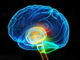
There is an endocrine gland in your brain called the pineal gland. Located deep within the brain, the gland is shaped like a tiny pinecone, which is how it got its name (“pine”-eel gland). The pineal gland is the least understood gland of the endocrine system and was the last part of the system to be researched.
Probably the first person to discover the pineal gland was Herophilos (325-280 B.C.), a famous anatomist at the University of Alexandria in Egypt. He was also the first to write about the soul being equated to the pineal gland. Fast forward 1,800 years to the Middle Ages when renowned anatomist Rene Descartes (1596-1650) also promoted the pineal gland as the seat of the soul. The theory that connected the pineal gland with man’s spiritual center remained part of the explanation of its function for nearly 2000 years.
It wasn’t until the late 1800s that the pineal gland began to undergo rudimentary study of its anatomy, histology, and embryological origins. The true function was not identified for nearly another 100 years, when Bargmann (1943) began to study the endocrine functions of the gland. The 1970s brought an acceleration of research through the advent of the electron microscope, which revealed its very complex anatomy, cytology (cell structure) and innervation, and its ability to interface or produce 5-HTP, serotonin, and melatonin.
An important part of the history of pineal research was the discovery of its relationship between light/dark periodicity and the body’s circadian rhythm. It has been discovered that the pineal gland interfaces with various by secreting indoleamine and polypeptides that interface with other segments in the brain in the following ways: (source)
Hypothalamus - generates vasopressin (blood pressure regulation) and oxytocin (effects organs of reproduction in men and women)
Pituitary gland - generates stimulatory hormones (FSH, LH, TSH, ACTH, etc)
Endocrine organ - parathyroid glands, pancreas
Regulates sleep - regulates the circadian sleep cycle by being the primary producer of melatonin
The pineal gland has a very rich network of blood vessels. The blood flow to the pineal gland is only superseded by the blood supply to the kidneys. Because it is not protected by the blood-brain barrier, the pineal gland may accumulate significant amounts of calcium, cobalt, zinc, selenium and fluoride.
As recently as 2020, a review article was published in the Journal of Applied Sciences entitled “Fluoride and the Pineal Gland.” Researchers noted that the pineal gland is most likely the most fluoride-saturated organ of the human body, noting that the range of recorded values was very wide but often similar or even higher than the levels observed in bones and teeth. The amount of fluoride in the gland exceeds the amount in other soft tissues, such as in muscles, by many times.
These authors (and others) believe that fluoride exposure may contribute to increased pineal gland calcification and which may lead to melatonin and other hormonal deficiencies, which in turn contributes to sleep disturbances.
Calcification of the pineal gland can also lead to Alzheimer’s disease, kidney disease, schizophrenia, and various forms of migraines.


